Types of Imaging
The Cancer Center of Southern California is a treatment and clinical research facility that is dedicated to providing the most comprehensive and technologically advanced cancer therapies available. We develop a personalized course of treatment for each patient using a variety of state-of-the-art imaging devices, such as those detailed below, in order to evaluate and monitor the extent of the patient’s cancer and treatment progress.
X-ray

Uses electromagnetic radiation to detect differences in materials or densities of tissues within the human body.
Computed Tomography (CT/CAT scan)
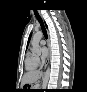
Diagnostic exam that uses X-rays to take cross-sectional images of the human body. Helps in tumor detection, staging cancer, and changes occurring in cancerous tissue due to treatments.
Magnetic Resonance Imaging (MRI)
Diagnostic exam which uses magnetic fields to create a detailed computer generated images of internal organs and tissues. Often used to diagnose and evaluate tumors in the chest, abdomen and brain.
Learn more about MRI scans from WebMD.com.
Mammogram
Type of X-ray specifically designed to view small tumors or irregular tissue within the breast.
Positron Electron Tomography (PET Scan)

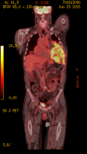
Diagnostic exam which uses a non-harmful radioactive tracer to detect disease within the body. The radioactive tracer is introduced into the blood stream via an IV and collects in a specific tissue type. The PET scanner detects and converts the radioactive tracer signal into 3D images that can be viewed by physicians.
Echocardiogram (EKG)
An imaging device which uses sound waves to create an image of the heart. It is used to check heart function before and during chemotherapy administration, and identify pre-existing heart conditions.
Ultrasound
A device which converts the echoes of high frequency sound waves to create images of internal organs. These echoes depend on the composition and density of tissue. Tumors have different densities and compositions in comparison to their surrounding tissue, and can thus be visualized by this method.
Bone Scan
A diagnostic test used in determining bone damage done through cancer, infection or trauma. An image is created by using a non-harmful radioactive tracer which enters the bones and is detected through the use of a special camera.
PET/CT Scan

PET and CT scan are performed together to provide extremely detailed images of tissues and organs within the body. Combining a PET scan which detects any abnormalities within the body, and the CT scan which shows a detailed map of the body, provides a complete image that neither test can offer alone.
Contact the Cancer Center to Learn More
The Cancer Center of Southern California is dedicated to providing the best treatments available for fighting cancer. Our team places each and every one of our patients’ physical and emotional well-being as a top priority and strive to ensure that our patients are comfortable and confident in our care. We offer a variety of services that combine medical oncology, hematology, and internal medicine. Please contact us at 310-552-9999 to schedule a consultation with one of our skilled Los Angeles oncologists.
Next, learn about Chemotherapy.


